Problems with using or understanding language (aphasia) appear, according to statistics from studies conducted by NABI (National Stroke Association), in 25 patients out of 100. Every fourth patient experiences difficulties associated with verbal communication - the ability to speak, write or understand spoken language. and written language. This indicates that acute cerebrovascular accident affected one or more language control centers.
Aphasia is a symptom of brain damage, not a disease. As individual as the picture of damage is, the manifestation of aphasia in each patient is as individual. This condition determines the individual nature of the selection of speech restoration techniques after a stroke for each patient.
Types of aphasia
Classification of sensory aphasia after stroke (Wernicke's disease):
1. Mnestic - characterized by the slowness of pronunciation of words (including those that are not suitable in context to the surrounding environment), the inability to concentrate on one thought in order to correctly formulate phrases. In addition, a person is able to remember only a couple of words from what he hears from his interlocutor. In turn, it includes the following subtypes: optical-mnestic, acoustic-mnestic.
2. Semantic - manifests itself in the difficulty of perceiving actions related to logical thinking, as well as long sentences.
3. Amnestic - the patient cannot remember the name of individual objects, but at the same time can describe what they are intended for. For example, “shop” is “a place where you can buy things/products”, etc.
Motor aphasia after stroke includes the following types:
1. Dynamic - manifests itself in the meaninglessness of phrases spoken by a person, which is provoked by an involuntary rearrangement of words in sentences. In this case, the patient is aware of the incorrect pronunciation, but cannot influence the situation in any way.
2. Afferent - manifests itself in the rearrangement of sounds in spoken words. For example, replacing voiced consonants with voiceless ones.
3. “Broca” - speech is pronounced with large intervals between syllables and words. It may manifest itself as frequent unintelligible repetition (like stuttering), mooing, etc.
The written speech of patients with this disorder is characterized by a large number of errors. Sometimes people start replacing words with opposite meanings. With total aphasia, there is simultaneous difficulty in understanding and pronunciation.
Sensorimotor (total) aphasia after stroke: prognosis
The mortality rate among hospitalized patients with speech defects exceeds the same figure compared to the post-stroke state, in which there are no problems in the perception and reproduction of speech. More detailed statistics on life prognosis can be found in this article.
Prediction of the effectiveness of rehabilitation therapy is carried out on an individual basis - the attending physician assesses the amount of damage and the affected area of the brain.
There are four main types of post-stroke speech disorders
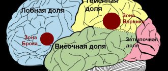
Damage to the language center in the dominant hemisphere - Broca's area - leads to the fact that patients lose the ability to convey their thoughts using coherent and grammatically correct language structures. Conversely, pathological changes in Wernicke's area (sensory center) lead to problems in receptivity to language. Such patients have difficulty understanding spoken or written language. They often use correct grammatical structures, but their statements may not make sense. The most severe form of aphasia (global or total) occurs in patients with extensive damage to several areas of the brain responsible for understanding and language. The mildest form of aphasia—amnestic—leads to difficulty working with vocabulary.
How do these types of aphasia manifest themselves in practice?
Damage to the language center in the dominant hemisphere - Broca's area - leads to the fact that patients lose the ability to convey their thoughts using coherent and grammatically correct language structures. Conversely, pathological changes in Wernicke's area (sensory center) lead to problems in receptivity to language. These patients have difficulty understanding spoken or written language. They often use correct grammatical structures, but their statements may not make sense. The most severe form of aphasia (global or total) occurs in patients with extensive damage to several areas of the brain responsible for understanding and language. The mildest form of aphasia—amnestic—leads to difficulty working with vocabulary.
How do these types of aphasia manifest themselves in practice?
- Motor aphasia. You know what you want to say, but you can’t find (remember, use and pronounce) the right words.
- Sensory aphasia. You hear someone speak or see printed text, but cannot understand the meaning of the words.
- Amnestic aphasia. You find it difficult to select and use the correct words to refer to certain people, objects, places or events.
- Global aphasia. You cannot speak, write, read and do not understand what is being said to you.
Exercises for aphasia after stroke
To quickly restore speech function, it is recommended to perform special exercises under the guidance of a speech therapist-aphasiologist. The sooner classes begin after a stroke, the more effective the activities will be.
It is recommended to begin classes immediately after transferring the patient to a regular hospital setting (with the exception of intensive care wards).
Training begins with light loads (up to 7 minutes per day), gradually increasing the time of interaction with a highly specialized specialist (up to 15 minutes/day). Note: the key role is not the duration, but the frequency and regularity of classes.
Basic exercises are built according to the standard scheme:
• reading; • written loads; • construction of proposals (occurs in the form of a confidential dialogue); • singing; • speech therapy massage - a specialist massages the cheeks, areas of the tongue, palate, lips using a disposable wooden or plastic otolaryngological spatula; • manipulations aimed at achieving the patient's understanding of what is happening around him and the sentences being spoken. Sometimes they resort to the use of gestures and facial expressions.
Effective loads for eliminating aphasia in stroke (speech therapy material for classes):
1. Curling your lips into a tube and trying to hold them in this position for at least five seconds. A similar exercise is done with the tongue. 2. Inflating the cheeks with air. 3. Alternately grabbing the upper and lower lips with your teeth. 4. Performing circular movements with the tongue clockwise and counterclockwise (so-called lip licking). 5. Imitation of a kiss. 6. It is necessary to reach the chin/nose with your tongue. 7. Tapping the tip of your tongue on the roof of your mouth. 8. Study and regular repetition of elementary words, incl. using data visualization (paper pictures, special mobile applications, PC programs, etc.). 9. Learning and honing the quality of speaking tongue twisters. Note: to control the quality of the exercises, it is recommended to perform the listed manipulations in front of a mirror. The lion's share of success depends on self-reinforcement of the material. We are talking about conducting training with the family and friends of a person who has suffered cerebral edema. The patient should be praised for any successful attempt.
What causes aphasia during a stroke?
In the human brain there are several interconnected centers responsible for oral speech: for its understanding, reproduction, analysis of complex speech structures, and the ability to construct correct sentences. All of them are interconnected by nerve fibers and are located mainly in the central part of the brain, as well as in the temporal and parietal lobes. Some of these speech centers are symmetrical in both hemispheres (that is, they are duplicated in each of them), but there are also areas that right-handers have only in the left hemisphere, and left-handers have in the right.
When a stroke occurs, part of the brain dies. If death occurs in one of the speech centers, or the nerve endings connecting these zones are damaged, aphasia develops. Thus, aphasia is a violation of the understanding or reproduction of already formed oral speech, sometimes up to its complete absence. If the disorder concerns written speech, then such a neurological syndrome will already have a different name (alexia, agraphia).
Treatment
Treatment of amnestic-semantic aphasia must be comprehensive. In this case, one should not hesitate, since the sooner corrective measures are started, the greater the patient’s chances of restoring lost cognitive functions.
After a diagnostic examination and clarification of the severity of damage to the speech centers of the brain, specialists formulate an individual treatment plan. In drawing up a treatment plan, in addition to functional and organic disorders, the patient’s age and mental state play an important role. Both a neurologist and a psychotherapist must work with each patient to achieve maximum effectiveness from therapy. If a serious organic pathology is detected that requires immediate surgical treatment, for example, in case of brain injuries or tumor lesions, emergency or planned surgical intervention is performed to eliminate the life-threatening condition and minimize cognitive impairment in the future.
Like a child!
Speech is a complex process in which different areas of the cerebral cortex actively interact. Thus, Wernicke's area is responsible for understanding; signals are formed in Broca's area, which ultimately turn into words and phrases. The role of auditory and visual analyzers is important. When a person talks, the whole brain is actively working. It establishes connections, for example, between the image of a spoon and the word that denotes this object. It is these connections that are destroyed in patients with amnestic aphasia. This is the difference between patients of the NeuroSpectrum Center for Pediatric Speech Neurology and Rehabilitation, who are being treated for amnestic aphasia, and healthy children. The normal process of forming connections goes by itself, and if connections are destroyed, their restoration requires outside help. Fortunately, children's brains are plastic, and it is easier to help our patients than adults and especially older people.
Motor aphasia
Broca's motor aphasia
is a violation of the ability to speak while maintaining the ability to understand speech. Occurs when the frontal lobe of the brain (Broca's motor speech center) is damaged. The patient has difficulty pronouncing or does not pronounce words, mainly pronouncing simple words or syllables. The main features of the form of motor aphasia are:
- the patient’s speech is poorly distinguishable, but is often understandable and meaningful, accompanied by eloquent gestures (the patient has difficulty pronouncing and moving from one word to another)
- the speech of others is well understood by the patient
Prognostic role of IS-related factors
A retrospective analysis of data from the acute period of stroke from the study cohort ( n
= 40) revealed a direct positive relationship between the initial severity of IS (NIHSS scores on the 1st day of IS) and the initial degree of aphasia on days 1-3 of IS (β = 0.08;
p
≤0.02), while the severity of IS (NIHSS scores on day 1) is an independent prognostic factor for the risk of severe aphasia on days 1–3 of IS (OR 4.07; 95% CI [1.25– 13.24]).
Similar results were obtained in a number of clinical studies of the acute period of stroke [35, 36]. The independent prognostic value of this indicator also remains for assessing the degree of aphasia by the 3rd month of AI (OR 3.27; 95% CI [1.02–9.77]). The degree of functional recovery of patients by the 21st day (mRs scores) is significantly positively associated with the recovery of speech functions by the 3rd month of AI (β=1.24; p
≤0.002), although this indicator is not an independent prognostic factor.
The initial severity of aphasia on the 1st day of IS is considered as an independent risk factor for the severity of aphasia by the 3rd month of IS and the 1st year of the disease [37, 38]. The results obtained do not confirm the independent prognostic significance of the initial severity of aphasia for the subsequent restoration of speech function by the 3rd month of the disease. At the same time, linear regression data revealed a direct relationship between the severity of aphasia on the 1st day and the degree of speech impairment by the 3rd month of the disease (β=1.2; p
≤0,000).
In the acute period of IS, 9 (22.5%) patients had a combination of aphasia and dysarthria syndromes. By the 3rd month of the disease, 6 (15%) of them had a regression of dysarthria while maintaining aphasia, and 3 (7.5%) had a regression of aphasia while maintaining dysarthria. In patients with a combination of aphasia and dysarthria in the acute period of IS, the likelihood of persistence of aphasic syndromes after 3 months is associated with a high degree of disability according to mRs ( p
<0, OR=0.31; 95% CI [0.27–0.35] [39].
Analysis of the statistical significance of the influence of focal motor and sensory neurological deficits in the early recovery period of the disease (total NIHSS score) on the severity of aphasia (average score on speech subtests of the 10-point scoring method) by the 3rd month of the disease before the course of therapy revealed a significant dependence of these signs ( p
=0.005, OR 4.6; 95% CI [1.39–15.11]). The distribution of the severity of speech disorders depending on the initial NIHSS value is presented in Fig. 3.
Rice. 3. Linear positive relationship between the degree of speech impairment and the severity of focal neurological deficit on the NIHSS scale (y-axis) upon admission of patients to the hospital. The x-axis shows scores on the speech subtests of the 10-point HPF assessment method; the y-axis shows NIHSS scores. A similar relationship was observed between the functional recovery of patients (total Barthel index score) and the degree of speech impairment ( p
= 0.004, OR 3.92; 95% CI [1.01–15.21]).
After a course of rehabilitation treatment, an independent effect on regression of aphasia was associated only with the severity of focal neurological deficit according to the total NIHSS score ( p
= 0.001, OR 6.4; 95% CI [1.75–23.35]).
Thus, the severity of motor and sensory deficits is an independent factor influencing the degree of recovery of speech functions by the 3rd month of the disease. The above data suggest that it is one of the pathogenetic factors that worsens the processes of post-stroke plasticity of neuronal networks.
According to neuropsychological examination, all patients showed a decrease in the severity of speech disorders. A significant improvement in speech function was observed in 6 (15%) patients with gross sensory function (by an average of 1.05 points - from 5.09 to 6.14 points according to the 10-point assessment method for HMF) and gross sensorimotor function (by an average of 1.05 points). 03 points - from 2.91 to 3.94 points) aphasia (Fig. 4).
Rice. 4. Dynamics of speech restoration after a course of complex rehabilitation therapy in patients with different forms of aphasia [22]. The abscissa axis shows forms of aphasia; the ordinate axis shows the dynamics of speech restoration, scores. Preserved speech function corresponds to a scale value of 10 points. The observed significant (by more than 1 point) regression of aphasia in patients with mixed and sensory types in all cases led to a change in the degree of its severity to a lighter one at discharge. This result is consistent with observations of maximum regression of sensory aphasia during directed speech therapy in the early recovery period of IS. In the late recovery period of IS, the highest effectiveness of speech therapy was revealed [40] for mixed sensorimotor aphasia.
Thus, the results of our study do not support the hypothesis that certain clinical forms of aphasia have an unfavorable prognostic significance on the effect of rehabilitation treatment. There was no unfavorable prognosis for sensory aphasia for regression of symptoms after a course of rehabilitation treatment. These results do not agree with the data of a number of authors [41–43] about the low therapeutic effectiveness and negative prognostic value of sensory aphasia. This issue probably requires further research. No statistically significant relationship was found between the degree of restoration of speech function and a specific form of aphasia.
A high rate of regression of non-speech disorders was observed in 32 (80%) patients with mixed and sensory (40% of cases each) and 8 (20%) with complex motor aphasia. In 70% of cases, significant regression according to the 10-point assessment method was combined with the presence of severe aphasia, in 40% - moderate. Insignificant dynamics of recovery of non-speech functions was observed in initially mild aphasia: 28 (71%) patients had mild to moderate speech impairment. Thus, in patients with severe speech disorders, cognitive rehabilitation is highly effective in restoring non-speech disorders.
A high significant positive correlation was found between the degree of non-speech impairment and the severity of aphasia ( r
=0.73;
p
<0.05) in patients under 65 years of age.
When calculating this indicator for the entire sample, a high significant positive correlation was also obtained, although the value of the correlation coefficient was lower ( r
= 0.64;
p
< 0.05).
It has been shown that as patients age, the severity of speech disorders becomes more related to the degree of neurodynamic impairment. Thus, in patients over 65 years of age, a significant high correlation was obtained between the degree of non-speech and neurodynamic disorders ( r
= 0.74;
p
< 0.05).
Depressive disorder, qualified as a single depressive episode of varying duration, was identified in 23 (57%) patients according to the psychiatrist’s conclusion and the results of testing according to IASOD-10 (total score ≥6). Upon admission to TsPRiN, the average score on IASOD-10 was M 15.5; 95% CI [6.6–24.4]. According to the regression analysis, a direct positive relationship was revealed between the severity of aphasia (according to the 10-point assessment method) and the development of post-stroke depression (total score ≥6 on the ASHOD-10; β=0.17; p
≤0.03). Determining the severity, clinical features of the course and typological distinctions of depressive disorders in patients who are not capable of verbally describing complaints and emotional state presents significant difficulties [44]. In these cases, it is impossible to use classical diagnostic methods associated with questionnaires using special scales or detailed interviews with patients. Therefore, for the initial identification of depression in the presence of speech dysfunction, it is necessary to use standardized observational scales that record the external manifestations of psychopathological symptoms and syndromes [45, 46]. Using screening using the Russian version of the hospital version of IASHOD-10 and examination by a psychiatrist, depressive disorder was identified in 57% of cases [19, 45]. The results of studies [47, 48], which used other observational scales, also indicate 44–70% of cases of anxiety and depressive disorders in post-stroke aphasia. The identified direct relationship between the severity of aphasia and psychopathological symptoms probably reflects the predominantly exogenous nature of depression. A direct relationship between the severity of focal neurological deficit and depression, showing the reactive nature of the latter, was also observed in the recovery period of IS in other clinical studies [49, 50].
According to a number of studies, the size and localization of post-infarction changes in the speech areas of the left hemisphere, as well as the basal ganglia, are unfavorable predictors of aphasia regression [5, 35, 51], while the localization of the lesion in the left superior temporal gyrus is associated with an unfavorable prognosis for the restoration of speech functions [52]. In contrast to the above data, in the present study, no reliable negative prognostic value was obtained for MRI signs associated with the size of the lesion, damage to various structures of the left perisylvian region and basal ganglia, however, an intact (according to structural MRI) area of the left superior temporal gyrus associated with good recovery of speech function.
The results obtained indicate that there is no direct dependence of the regression of aphasia on the localization of the focus in the early recovery period of I.I. This is likely due to significant variability in the recovery or compensation of speech function associated with the reorganization of intra- and interhemispheric speech neuronal networks. This is consistent with previously obtained data [53] that traditional MRI predictors of regression of post-stroke aphasia in the acute period of IS are insignificant for the later (90 days of IS) period of the disease.
In contrast to the regression of aphasia syndrome, a number of MRI signs showed a significant relationship with the dynamics of non-speech cognitive impairment. A positive linear relationship was revealed between lesions of the left angular gyrus (β=0.92; p
≤0.01) and the absence of regression of non-speech cognitive impairment.
A negative linear relationship was obtained between the localization of post-stroke changes in the left frontotemporal region (β= –0.6; p
≤0.05) and regression of non-speech cognitive, while the volume of the lesion turned out to be significant (β=0.002;
p
≤0.000).
It is known that the leading cause of the formation of chronic cerebrovascular accident is damage to small-caliber arteries caused by hypertension, atherosclerosis, diabetes and their combination [54]. In the study cohort, 80% of patients had MRI signs of varying severity of microangiopathy, probably caused by this underlying vascular pathology. However, according to logistic regression, the severity of MRI signs of microangiopathy is not associated with regression of aphasia (β= –0.19; p
≤0.39) and non-dementia cognitive impairment (β= –0.06;
p
≤0.95).
Modifiable vascular risk factors for IS and aphasia prognosis
According to logistic regression, there was no significant influence of vascular risk factors for IS on the regression of aphasia. Probably, the absence of a direct pathogenetic connection in this case is due to the complex individual functional reorganization of the speech structures of the brain caused by neuroplasticity [55].
According to the results of prospective clinical studies [56, 57], a number of cardiovascular risk factors for IS, including hypertension, type 2 diabetes, hyperlipidemia, are also risk factors for the progression of non-dementia cognitive impairment and the formation of vascular dementia. Our patients revealed a significant linear relationship between the presence of type 2 diabetes (β=0.06; p
≤0.02), hyperlipidemia (β=0.11;
p
≤0.000) and the degree of non-speech (non-dementia) cognitive impairment using a 10-point assessment method. This cause-and-effect relationship confirms the significance of vascular risk factors associated with the pathology of intracerebral arteries and microvasculature for impaired autoregulation of cerebral blood flow, one of the pathogenetic mechanisms of cognitive disorders [58].
Thus, to optimize the process of rehabilitation therapy and develop new approaches to neurorehabilitation of patients with post-stroke aphasia, it is necessary to take into account prognostic factors that influence the regression of speech and non-speech cognitive impairment. A number of risk factors associated with I.I. have been identified. For regression of aphasia both in the acute and recovery periods of the disease, the initial severity of neurological symptoms (total NIHSS score on days 1–3 of stroke) has an independent prognostic value. At the 3rd month of the disease, the independent effect on regression of aphasia is associated with the severity of focal neurological deficit (NIHSS score) and the degree of functional recovery (Barthel index). A number of MRI signs (localization of post-stroke changes in the left angular gyrus, frontotemporal region and lesion volume) had a significant effect on the dynamics of non-speech cognitive impairment. At the same time, traditional MRI predictors of regression of post-stroke aphasia in the acute period of IS turned out to be insignificant for the recovery period of the disease. Therefore, the search for other significant prognostic factors must continue. Biological and social factors (age, gender, education) do not have independent prognostic significance, although a direct relationship between age and certain clinical forms of aphasia has been shown, as well as a significant influence of higher education on the regression of aphasia. Neuropsychological data indicate the influence of its severity on the degree of improvement in speech function. A significant improvement in speech function was revealed in patients with initially severe forms of aphasia, in particular sensory and sensorimotor. In these cases, the effect of speech therapy led to a change in the severity of the disorders. The study results indicate a high level of depression in patients with aphasia. A direct relationship has been revealed between depression and the severity of speech disorders. Considering the negative impact of depression on the rehabilitation process, continued research is necessary for the early diagnosis of affective disorders in this category of patients.
This work was supported by the Russian Science Foundation grant No. 16−15−10419.
The authors declare no conflict of interest.
Symptoms and manifestations
Amnestic aphasia is characterized by a mild course of the disease and a blurred manifestation of the clinical manifestations of the disease. Among the most common manifestations are the following symptoms:
- Slow speech of the victim, with pronounced intervals between sentences, or vice versa, the victim’s speech becomes fluent, illogical, turning into “porridge”;
- Repeated repetition of the same words or phrases;
- The presence of paraphrases is a descriptive characteristic of the semantic load of a word. Speech becomes descriptive;
- The patient misses words, most often nouns;
- Also, the patient cannot remember the name of the item or object in question, but knows its function and appearance.
Despite this, the patient's reading and writing skills do not suffer. His speech is constructed correctly, both grammatically and logically. The patient does not have difficulties with the acoustic pronunciation of words, as well as articulation. The patient's speech becomes expressive and replete with a large number of verbs.
It is important to note that the patient easily remembers the name of an object when prompted or in context, for example, if you name the first syllable of the desired word, the patient will easily remember its ending.
Symptoms of aphasia
Signs and symptoms of aphasia may be invisible to others, as compensatory processes are activated that conceal speech defects. In the early stages of the disease, symptoms may appear or disappear completely, so it is difficult to draw a clear line between impaired and normal speech. Aphasia is a systemic disorder in which not only the pronunciation side suffers, but also the understanding of speech, lexical and grammatical structure, reading and writing. Symptoms of aphasia also manifest themselves in neurological and mental abnormalities. Motor functions suffer, patients experience paralysis of the limbs and articulatory muscles. Voluntary movements acquired throughout life experience are impaired. So the patient seems to forget how to use cutlery and household items. Memory and attention are severely impaired, and thought processes are inhibited.
A.R. Luria identifies six main forms of aphasia: acoustic-gnostic (sensory), acoustic-mnestic, afferent motor, efferent motor, semantic and dynamic.
Up
Who destroyed the bridge in the head?
Typically, a particular type of speech disorder is associated with problems in the functioning of a specific area of the cerebral cortex, and this helps doctors make a diagnosis. But not in this case. In both left-handers and right-handers, amnestic aphasia develops most often with damage to:
1) basal or anterior parts of the temporal lobe,
2) temporo-parietal-occipital junctions,
3) inferior parietal lobule.
Foci are also found in other areas of the cerebral cortex. The causes of violations are various. In children, in most cases, amnestic aphasia occurs when the brain is damaged as a result of trauma, but the disease also occurs in those who have experienced severe poisoning with brain damage, neuroinfection, or suffer from epilepsy and tumor diseases of the brain. But the essence is always the same - the transferred condition led to the death of neurons in a certain area of the cortex. Speech is the result of complex brain work. Therefore, amnestic aphasia is observed in a wide variety of lesions.
Why look for a reason?
Amnestic aphasia is only a symptom of severe brain damage. To successfully fight it, you need to make sure that the patient does not get worse. Therefore, neurologists are actively involved in the treatment of the disease. If the cause is not removed, the localization of damage will expand, and amnestic aphasia will be combined with other disorders. Often this is facial agnosia, in which the patient cannot even recognize relatives, not only in photos, but also in life.
Therefore, patients undergo various studies. Computed tomography or magnetic resonance imaging will help determine if there are any abnormal changes inside the skull. In this way, it is not difficult to detect tumors, bone fragments after traumatic brain injuries, areas of tissue that are left without blood supply, and much more. Electroencephalography - recording the electrical signals that the brain sends - will help determine whether there are areas of hidden excitation of the cortex, that is, whether forgetting words is associated with signs of epilepsy. This insidious disease does not necessarily manifest itself in large convulsive seizures; there are also forms with subtle manifestations.
Examination by an experienced neurologist is extremely important. Our center’s specialists work a lot with speech problems in children. They know how to find an approach to any child, help him feel at ease, and sometimes notice signs that usually remain hidden from the eyes of the doctor, not only in the clinic, but also in the hospital.
